SonoBox: A Robotic Ultrasound System for Pediatric Forearm Fracture Diagnosis
Project Description
Pediatric forearm fractures are a common reason for medical visits and often require X-rays for diagnosis, which involve ionizing radiation that can be harmful to children. This is especially concerning because children's tissues are more sensitive to radiation than those of adults. This project explores ultrasound imaging as a safer and non-ionizing alternative through the development of the SonoBox system. We aim to replace radiation-based methods with a child-friendly solution that uses ultrasound technology.
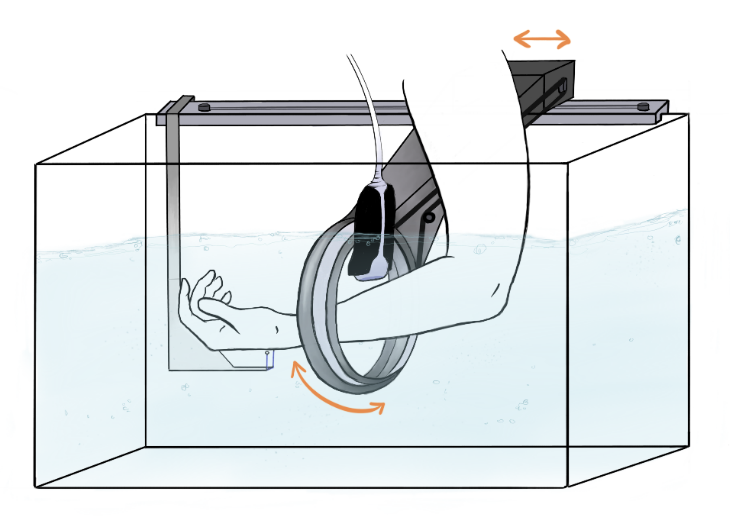
We are building an ultrasound-based, contact-free, and automated cross-sectional imaging system for diagnosing pediatric forearm fractures. A commercially available ultrasound probe is guided around the forearm in a water-filled tank using robotic control and utilizes image processing methods to generate a comprehensive ultrasound tomogram. Safety and hygiene considerations, gender and diversity relevance, and the potential reduction of radiation exposure and examination pain are pivotal aspects of this endeavor.
Preliminary experiments show that clear ultrasound images can be generated quickly in a water bath. The system overcomes issues like water turbulence during probe movement and can accurately track the probe position. The SonoBox prototype has shown promising results in transmitting position data for ultrasound imaging, indicating potential for autonomous, accurate, and potentially painless fracture diagnosis. The project outlines further goals, including the construction of prototypes, validation through patient studies, and development of a hygiene concept for clinical application.
The SonoBox project marks an important advance in diagnosing pediatric fractures and offers a safer and more comfortable alternative to traditional X-ray imaging. By automating the imaging process and eliminating the need for direct contact, SonoBox can improve clinical efficiency and reduce discomfort for young patients. It also opens new possibilities for using ultrasound in areas where it was previously limited. Ongoing research will use the current prototype as a foundation for a new design that prioritises user-friendliness and patient comfort, while also validating its performance in clinical settings and exploring its potential in other medical and veterinary fields.

- Research
- SonoBox: A Robotic Ultrasound System for Pediatric Forearm Fracture Diagnosis
- Robotics Laboratory (RobLab)
- OLRIM
- MIRANA
- Robotik auf der digitalen Weide
- KRIBL
- Ultrasound Guided Radiation Therapy
- Digitaler Superzwilling: Projekt TWIN-WIN
- - Finished Projects -
Floris Ernst

Gebäude 64
,
Raum 97
floris.ernst(at)uni-luebeck.de
+49 451 31015200
Rucha Golwalkar
Gebäude 64
,
Raum 83
rucha.golwalkar(at)uni-luebeck.de
+49 451 31015227