OCT-based Neuro-Imaging
Project Description
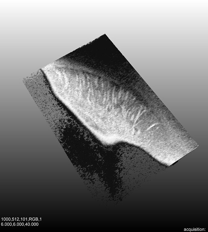
Optical Coherence Tomography (OCT) is a relatively young imaging technique. Based on the principle of interferometry, its functionality resembles the principle of ultrasound. In OCT imaging, however, near-infrared light with a wavelenght of 800 to 1500 nm is used which is scattered by the examined sample. The advantages of OCT imaging is the high resolution (1 – 10 mm), real-time capability, and the atraumatic imaging (no direct contact to the sample is needed). Disadvantage is the small imaging depth of 2-3 mm. OCT has been used in many medical applications, major developments have been made in the field of ophthalmology, dermatology, and urology. Little work has been done in the field brain imaging with OCT. It has been shown that tissue structure in vitro can be displayed by OCT (e.g. grey and white matter) and that tumorous tissue can be reliably identified.
From this state of the art, different research topics arise:
OCT for brain imaging
Despite the advantages of OCT, no explicit integration of OCT into the neurosurgical setup such as neuroimaging and neuronavigation has been done. As OCT could be integrated into minimal-invasive instruments, this kind of applications would be supported. Such an integration would for example allow for a real-time validation of the planned path. Several goals are focused on: development of a minimal-invasive probe for neuroimaging of the deep brain, development of brain tissue classification algorithms, and integration into the neuronavigation.
Speckle reduction in OCT images
As ultrasound images, OCT images are subject to speckle noise. Although speckle contains an information carrying part, this kind of noise often distorts the image and complicate subsequent use for diagnosis. In this project, new methods of speckle noise reduction are to be developed. This will be done based on two approaches:
(a) development of novel image processing methods and
(b) modified imaging modalities.
Tissue structure identification in OCT images
Closely related to the speckle noise reduction is the task of information extraction in OCT images. For diagnostic purposes (such as layer identification in the retina), the extraction of tissue structures is often of major interest. This project aims at the development of novel tissue structure identification methods in OCT images supporting the diagnosis or the orienation of the surgeon.
Publications
2008
Towards Multi-Directional OCT for Speckle Noise Reduction, Metaxas, Dimitris and Axel, Leon and Fichtinger, Gabor and Székely, Gábor, Eds. New York, NY, USA , 2008. pp. 815-823.
| DOI: | 10.1007/978-3-540-85988-8 |
| File: | 978-3-540-85988-8 |
2007
Model-Based Detection of White Matter in Optical Coherence Tomography Data, Lyon, France , 2007.
Towards Automated OCT-based Identification of White Brain Matter, 2007.
| DOI: | 10.1007/978-3-540-71091-2 |
| File: | 978-3-540-71091-2 |
2006
3D Visualization and Identification of Rat Brain Structures using Optical Coherence Tomography, Hannover , 2006.

- Research
- SonoBox: A Robotic Ultrasound System for Pediatric Forearm Fracture Diagnosis
- Robotics Laboratory (RobLab)
- OLRIM
- MIRANA
- Robotik auf der digitalen Weide
- KRIBL
- Ultrasound Guided Radiation Therapy
- Digitaler Superzwilling: Projekt TWIN-WIN
- - Finished Projects -
- High-Accuracy Head Tracking
- Neurological Modelling
- Modelling of Cardiac Motion
- Motion Compensation in Radiotherapy
- Navigation and Visualisation in Endovascular Aortic Repair (Nav EVAR)
- Autonome Elektrofahrzeuge als urbane Lieferanten
- Goal-based Open ended Autonomous Learning
- Transcranial Electrical Stimulation
- Treatment Planning
- Transcranial Magnetic Stimulation
- Navigation in Liver Surgery
- Stereotactic Micronavigation
- Surgical Microscope
- Interactive C-Arm
- OCT-based Neuro-Imaging
Achim Schweikard

Gebäude 64
,
Raum 94
achim.schweikard(at)uni-luebeck.de
+49 451 31015232
Floris Ernst

Gebäude 64
,
Raum 97
floris.ernst(at)uni-luebeck.de
+49 451 31015200
Ehemalige Projektmitarbeiter
- Dr.-Ing. Lukas Ramrath